
Artificial intelligence is changing the way people in every field work, including ophthalmology. Launched in 2024, Northwestern’s Center for Engineering in Vision and Ophthalmology (CEVO) is using AI and other cutting-edge technology to advance its glaucoma research. Backed by a gift from the Forsythe Family Foundation, CEVO is developing innovative imaging tools to better understand and treat glaucoma, one of the world’s leading causes of permanent blindness.
“The integration of engineering and AI is key to the future of medical diagnostics and interventions,” says Hao F. Zhang, co-director of CEVO and a professor of biomedical engineering at the McCormick School of Engineering. “The foundation’s support will bring the latest and most powerful AI hardware to CEVO to help us develop, train and apply new AI models and agents to achieve, for example, imaging-guided personalized surgeries.”
Glaucoma is a group of eye diseases that cause damage to the optic nerve. Under healthy circumstances, fluid in the eye called aqueous humor drains through a network of sophisticated tiny channels. When these pathways become blocked or don’t work properly, fluid builds up and intraocular pressure increases. Over time, this elevated pressure can lead to vision loss.
Current treatments include medication, which can become less effective over time. Minimally invasive glaucoma surgery, which lowers intraocular pressure by placing a stent along the drainage pathway, can be effective but has had limited overall success.
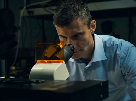
CEVO engineers are building new high-resolution imaging devices that capture the fine details of how fluid drains from the eye. These images, taken at a resolution of approximately 1 micrometer, are used to generate a digital twin of each patient’s eye.
Such high-resolution, AI-integrated imagery could enable more effective treatments for glaucoma by providing personalized information about each patient’s eye structure and function, allowing for tailored surgical planning. Instead of placing stents in the easiest insertion area, a surgeon could view a 3D digital model of the patient’s eye before surgery and know exactly where to place the implant for maximum efficacy.
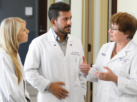
Nicholas J. Volpe, the George W. and Edwina S. Tarry Professor of Ophthalmology and chair of the department of ophthalmology at Northwestern’s Feinberg School of Medicine, co-directs CEVO with Zhang. Cheng Sun, a professor of mechanical engineering, and Mark Johnson, a professor of biomedical engineering, are also contributing to the research.
“One of Northwestern’s great strengths is the expertise it has in both engineering and ophthalmology,” Volpe says. “This gift from the Forsythe Foundation is perfectly timed to accelerate our discoveries, which will undoubtedly lead to better treatment for our patients.”
Reader Responses
No one has commented on this page yet.
Submit a Response